📝 Table of Contents
- Why Looking at Piles Images Matters for Diagnosis
- A Detailed Breakdown of Piles Stages with Visual Descriptions
- Stage 1 Piles Images: The Invisible Beginning
- Stage 2 Piles Images: The Prolapsing Phase
- Stage 3 Piles Images: Manual Reduction Required
- Stage 4 Piles Images: Permanent Prolapse
- Internal vs. External Piles Images: Spotting the Difference
- The Invisible Internal Piles
Piles Images Explained: A Visual Guide to Understanding Every Hemorrhoid Stage
Let’s be honest; nobody wakes up excited to search the internet for piles images. It is an uncomfortable, often embarrassing topic that many people hesitate to discuss even with their closest doctors. However, when you start noticing discomfort "down there"—itching, pain, or perhaps a frightening spot of blood on the toilet paper—seeking out piles images for comparison is usually the very first step people take in trying to figure out what is wrong. We search for these visuals because we want reassurance, understanding, and a way to gauge the severity of our own condition against a standard. But the internet can be a confusing place, filled with graphic photos that might not match what you are feeling. This guide is designed to cut through the confusion. We will provide a detailed, humanized walkthrough of hemorrhoids, describing exactly what medical diagrams and clinical piles images depict at every stage of development, helping you understand the symptoms and variations of this common condition.Why Looking at Piles Images Matters for Diagnosis
While self-diagnosis via Google images is never a replacement for a doctor's visit, understanding what you are looking at is crucial for your own peace of mind. Hemorrhoids (commonly called piles) are essentially swollen veins in your lower rectum and anus, similar to varicose veins you might see on legs. When doctors look at clinical piles images, they aren't just looking at a "lump." They are assessing location (internal vs. external), size, inflammation, and the degree of prolapse (how far they have fallen out of their normal position). For the average person, searching for piles images is about answering three questions: Is this what I have? How bad is it? And will it go away on its own? To answer these, we need to break down the condition stage by stage.A Detailed Breakdown of Piles Stages with Visual Descriptions
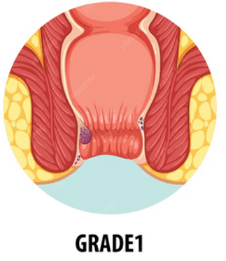
Doctors typically grade internal hemorrhoids on a scale of one to four based on their severity and whether they prolapse (come out of the anus). It is important to note that external hemorrhoids don't follow this same staging system, which we will discuss later. Below, we walk through the four stages. For each stage, we have provided a space for a corresponding visual representation. When looking at medical piles images online or in pamphlets, these are the distinct characteristics you should look for.Stage 1 Piles Images: The Invisible Beginning
Stage 1 is the mildest form of internal hemorrhoids. At this point, the veins inside the rectum have become swollen, but they remain securely inside. The Symptoms: Often, Stage 1 piles are asymptomatic, meaning you might not even know you have them. If symptoms do occur, they are usually mild:- Painless, bright red bleeding during bowel movements (you might see a small amount on the toilet tissue).
- Minor itching or irritation around the anal opening due to mucus discharge from the swollen internal tissue.

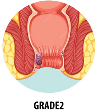
Stage 2 Piles Images: The Prolapsing Phase
Stage 2 is when the connective tissue holding the hemorrhoids in place begins to weaken. The key characteristic here is temporary prolapse. The Symptoms: The symptoms become more noticeable in Stage 2:- You may feel a lump or tissue coming out of the anus while straining during a bowel movement.
- Increased bleeding compared to Stage 1.
- A sensation of fullness or that the bowel hasn't completely emptied.
- Itching becomes more frequent due to the tissue irritating the sensitive skin around the anus.

Stage 3 Piles Images: Manual Reduction Required
By Stage 3, the condition has progressed significantly. The supporting tissues are stretched thin, and the hemorrhoids prolapse easily and frequently. The Symptoms: This stage is often painful and significantly affects daily quality of life:- The hemorrhoidal tissue prolapses during bowel movements, or even during physical activities like lifting heavy objects, coughing, or sneezing.
- Crucially, the tissue does not go back in on its own. You must gently push it back inside (manual reduction) with a finger.
- Pain, discomfort, and heavy itching are common.
- There may be discharge of mucus or fecal matter on underwear because the anal sphincter cannot close tightly around the protruding tissue.
Stage 4 Piles Images: Permanent Prolapse
Stage 4 is the most severe form of internal hemorrhoids. This is often considered a medical urgency, though not always an emergency unless blood supply is cut off. The Symptoms:- The hemorrhoids are permanently prolapsed. No amount of gentle pushing will get them back inside.
- Significant pain and discomfort, making sitting or walking difficult.
- High risk of thrombosis (blood clots forming inside the external hemorrhoid) or strangulation (blood supply cut off to the internal hemorrhoid), both of which cause excruciating pain.
- Constant bleeding and discharge.
Internal vs. External Piles Images: Spotting the Difference
When searching online for piles images, what you most commonly see are actually external hemorrhoids, or internal hemorrhoids that have reached Stage 3 or 4. It is vital to understand the difference, as the visual cues vary greatly.The Invisible Internal Piles
As discussed in the staging above, internal piles originate deep inside the rectum where there are few pain-sensing nerves. You usually cannot see them in standard piles images unless they have prolapsed. Their primary signal is bright red blood.The Visible External Piles
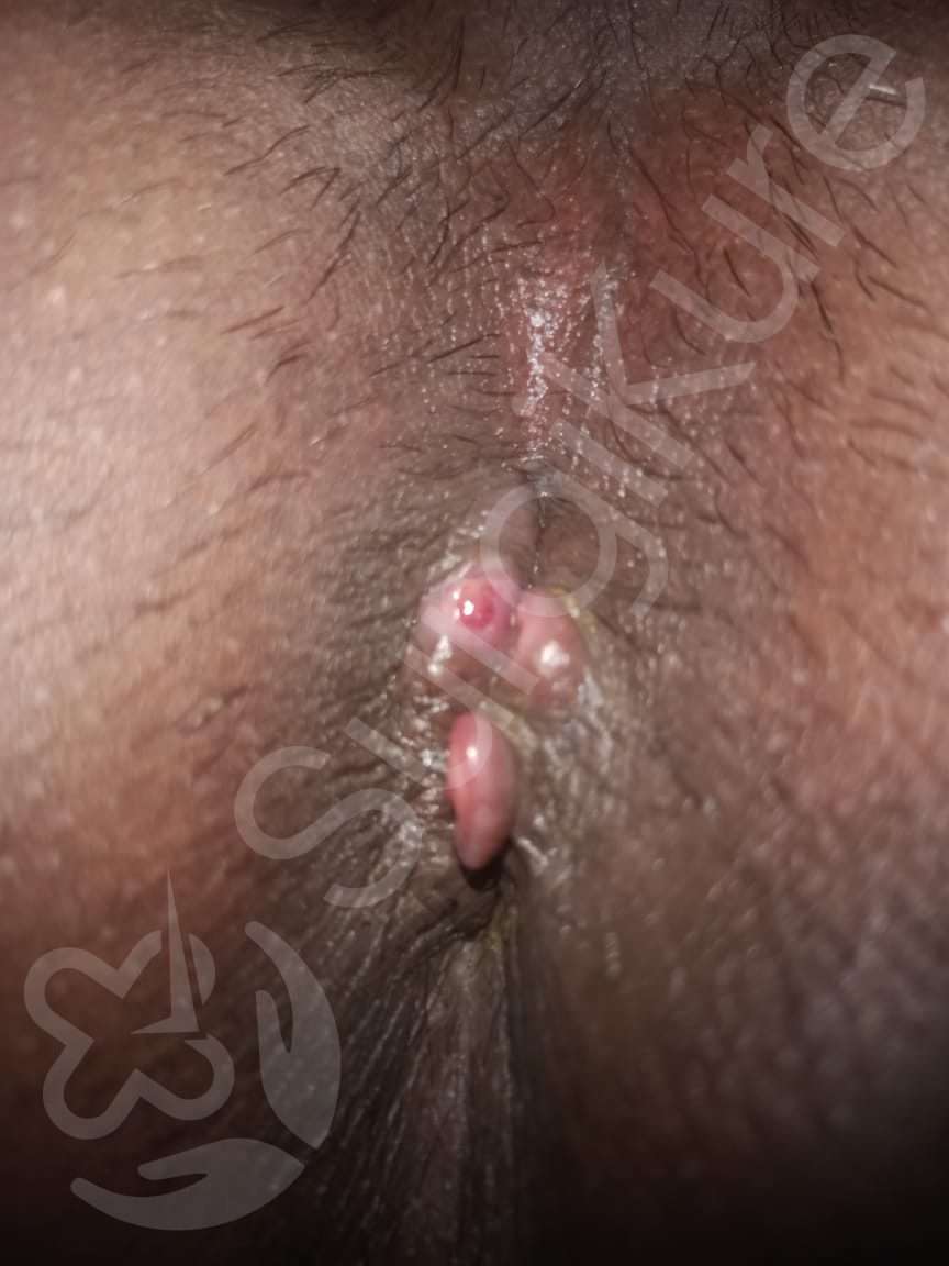
External hemorrhoids form under the skin around the anus, where there are many pain receptors. Describing External Piles Images: Visuals of external piles do not follow the Stage 1-4 progression. Instead, piles images of external hemorrhoids show visible lumps or bulges around the anal rim.- Normally: They may just look like extra folds of skin that feel itchy.
- Thrombosed: This is what leads to the most alarming piles images. If a blood clot forms in an external hemorrhoid, it turns into a hard, incredibly painful, bluish or purple lump the size of a pea or a grape. This appears suddenly and is very tender to the touch.
When to Move Beyond Piles Images and See a Doctor
We live in an era where we try to solve everything with a search engine. While looking at piles images can help you understand the geography of your problem, it is not a diagnosis. Many other conditions can mimic the appearance of hemorrhoids in online images, including anal fissures (tears), fistulas, skin tags, and, more seriously, colorectal cancer. You must move beyond just looking at piles images and seek professional care if:- You are experiencing bleeding for the first time. You must always get rectal bleeding checked to rule out serious causes.
- The pain is severe or came on suddenly (suggesting a thrombosis).
- Your hemorrhoids are at Stage 3 or Stage 4 (they won't go back in).
- Over-the-counter creams and fiber supplements haven't helped after a week.
- You have a family history of colon cancer.
Summary and Final Thoughts on Managing Piles
Navigating the world of medical information and trying to match your symptoms to graphic piles images on the screen can be an overwhelming and anxious experience. It is important to remember that you are not alone; hemorrhoids are incredibly common, affecting millions of people. By understanding the four stages of internal piles and the distinct appearance of external piles, you can better interpret the visual information you find. Remember that Stage 1 and 2 often respond well to lifestyle changes like increasing fiber intake, drinking more water, and avoiding straining. Stage 3 and 4 usually require medical intervention, ranging from minimally invasive office procedures (like rubber band ligation) to surgery in severe cases. Use the piles images you find as educational tools to empower yourself to seek help, rather than a source of fear or definitive self-diagnosis. The sooner you consult a doctor, the sooner you can move past the discomfort and get back to living your life.Stop Googling your symptoms and start treating them! At SurgiKure, your first step is on us. Get a 100% FREE Expert Consultation today: Call/WhatsApp: +91 7670968977
Ready to consult a specialist?
Free first consultation at SurgiKure. Same-day appointments available.